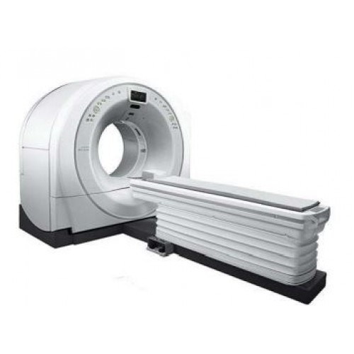
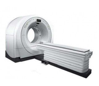
Supria 64/128 computed tomography scanner

- Warranty 12-24 months
- Warranty free service and post-warranty
- Equipment instalation and staff training at the company's expense
- Full customer support (phone consultation even after installation)
- Visiting the exhibition hall
- Leasing sale
- Holding conferences and events
- Quick response to a request
Supria Advantages
Patient-friendly and easy to fit into the healthcare facility. 75 cm gantry opening. The Speedia CT has one of the largest gantry openings in its class, reducing patient anxiety while occupying a compact footprint to facilitate CT installation in existing rooms.
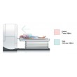
TILT ±30 °
The wide gantry tilt angle reduces artifacts from filled teeth and radiation dose to the eyes.
The large opening diameter (75 cm) of the Speedia provides easy access to the patient, even when the patient’s arms are raised or when the patient cannot lie on his back. This improves the work of the operating physician and the patient’s condition
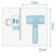
Using only the main system modules*: gantry, patient table and control console, Speedia has an impressively compact design. *Depending on the country, a system transformer may be required
High rotation speed during scanning High-speed scanning at a speed of less than 1 rpm. and advanced 3D reconstruction provide fast coverage for efficient and accurate examination.
Breathing Guide Three LCD displays inside the gantry port provide patient breath hold notifications, which, combined with automatic voice prompts, allow the patient to easily follow breathing instructions. ►lntelliEC - Automatic 3D mA modulation for dose optimization. The tube current is optimized in three directions (X-Y-Z) based on patient size information obtained from the scan and a given target SD. This allows for images with a constant noise level across the entire scan area, optimizing the balance between image quality and exposure. ►lntelli IR - Iterative processing for noise reduction. Advanced noise reduction technology using iterative reconstruction technology reduces noise and artifacts while maintaining high image quality at lower radiation doses
Software Description
Data Display: Patient name, birthday, gender, patient ID number, slice thickness, tube voltage, tube current, slice position, contrast status, and more.
Image Display Process:
(a) Multi-frame display.
(b) Magnification (real-time, etc.).
(c) Image rotation.
(d) Left-right inversion.
(e) Correction.
(f) Comment display.
(g) Filming.
(h) Subtraction/addition.
(i) Edge enhancement/smoothing.
(j) Multi-slice image addition.
Image Analysis Process:
(a) Distance and angle measurement.
(b) CT value profile.
(c) ROI setting.
- Shape: Oval, Free.
- Process: Area, CT Average #.
- Display: Up to 4 ROIs can be displayed.
- Control: Size, Position, Rotation.
(d) Histogram.
(e) CT value display.
(f) Scale display.
(g) Volume calculation.
3D image display
(a) MPR (SAG, COR, OBL, CURVE, SPINE).
(b) MIP, MinIP, RaySum display.
(c) Surface visualization.
(d) Volume visualization (VT method).
(e) Multi-angle reconstruction plan (MARP).
(f) Filming.
(g) Perspective method
Supply set:
1. Gantry scanner.
2. Patient table with examination accessories.
3. Operator console for data acquisition/processing with 24-inch LCD monitor.
4. Software package


