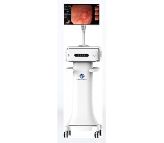
Video endoscopic system VC-880 (FHD)

- Warranty 12-24 months
- Warranty free service and post-warranty
- Equipment instalation and staff training at the company's expense
- Full customer support (phone consultation even after installation)
- Visiting the exhibition hall
- Leasing sale
- Holding conferences and events
- Quick response to a request
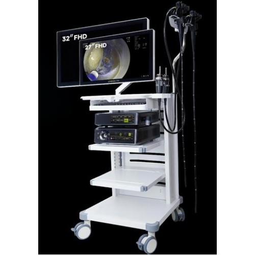
Video endoscopic system VC-880 (FHD)
Modern medicine requires not only highly qualified personnel, but also the use of innovative equipment capable of taking diagnostics and treatment to a fundamentally new level. These are the characteristics of the VC-880 (FHD) video endoscopic system, created taking into account the requirements of clinicians and the latest advances in visualization. Thanks to Full HD technology, intelligent software and ergonomic design, this multifunctional complex provides maximum accuracy with minimal time and effort on the part of the doctor.
Technical features and advantages
The VC-880 (FHD) video endoscopic system is the result of combining advanced engineering solutions, practical experience of medical specialists and modern IT developments. The complex has the following advantages:
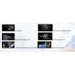
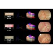
- High quality Full HD images. Thanks to the matrix with a Full HD resolution, a clear and contrasting image is obtained even in difficult clinical conditions. This is especially important in diagnosing early stages of diseases, where visual signs may be minimal.
- Artificial intelligence. The integration of AI technology into the VC-880 software allows real-time image analysis, recognition of abnormal areas and provision of preliminary conclusions. This function not only facilitates the doctor's work, but also significantly increases the overall efficiency of diagnostics.
- Ergonomics and ease of use. The video endoscopy system has a compact size, a convenient control panel, a touch screen and ample opportunities for individual parameter adjustment. All this makes working with the device as comfortable as possible even during long procedures.
- Flexibility in use. The system is compatible with a wide range of endoscopes and accessories, making it ideal for multidisciplinary clinics. Quick change of endoscopes and adaptive connectors provide versatility of use.
- Cost-effectiveness and reliability. The VC-880 has low energy consumption and high resistance to external influences. The design of the device is designed for intensive use, which means it will serve your clinic for many years without unnecessary maintenance costs.
At the same time, the price of the system remains very competitive. Current prices in Ukraine can be clarified with the specialists of "Rad Farm". Consultants will select the optimal configuration taking into account the budget and objectives of the medical institution.
Scope of application
Thanks to its wide functionality and high visualization accuracy, the VC-880 (FHD) video endoscopic complex is actively used in a variety of areas of medicine:
- gastroenterology - for esophagogastroduodenoscopy, colonoscopy and other gastrointestinal examination procedures;
- pulmonology - for bronchoscopy and examination of the tracheobronchial tree;
- surgery - used as a navigation tool during minimally invasive surgeries;
- urology and gynecology - for intracavitary diagnostics and monitoring of treatment effectiveness;
- otolaryngology - for examination of the nasal cavity, larynx, middle ear;
- pediatrics and neonatology - with the possibility of soft and safe visualization in children;
- veterinary medicine - used in clinics for animal diagnostics.
Such a wide range of applications makes the VC-880 video endoscope system a universal solution for both specialized offices and multidisciplinary medical centers. Moreover, the affordable cost on the Ukrainian market allows this technology to be implemented even in institutions with limited funding.
Why is it worth buying this device from Rad Farm?
Rad Farm is a reliable supplier of advanced solutions in the field of medical imaging. Our experience, reputation and professional approach make us the best choice for those who want to buy an innovative VC-880 video endoscope in Ukraine. We offer our clients the following:
- Official supply and certification.
- Favorable prices and transparent policy.
- Consultations and support at all stages.
- Support and service.
We promptly respond to requests, provide spare parts and update software, including components with artificial intelligence, which is especially important in the rapidly developing medical industry.
Make a choice in favor of efficiency — with VC-880 (FHD) from «Rad Farm»
If you are looking for a modern, reliable and affordable video endoscopy system in Ukraine, VC-880 (FHD) is the perfect choice. The complex combines the latest technologies, advanced functionality and ease of use. By contacting «Rad Farm», you get not just a device, but a complete solution for improving the level of diagnostics and treatment.
Contact our consultants to order or buy a modern VC-880 video endoscope, find out current prices, delivery times and possible configuration options. We will help you make the right choice and bring your institution to a new level of medical imaging.


-320x280.jpg)
