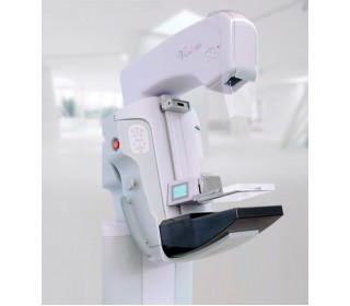
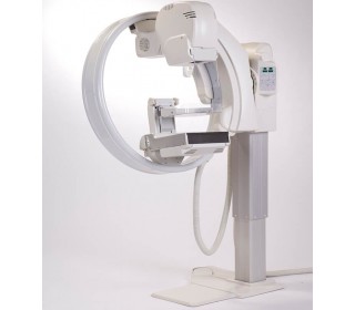
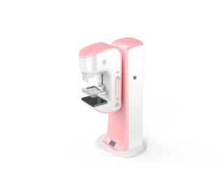
AMULET Digital Mammography System Innovality

- Warranty 12-24 months
- Warranty free service and post-warranty
- Equipment instalation and staff training at the company's expense
- Full customer support (phone consultation even after installation)
- Visiting the exhibition hall
- Leasing sale
- Holding conferences and events
- Quick response to a request
Advantages of the AMULET Innovality mammography system
1. The use of a high-efficiency flat-panel detector (FPD) ensures maximum electrical signal acquisition from X-rays to achieve high resolution (pixel size - 50 μm) at a low radiation dose.
2. The short interval between the end of one exposure and the start of the next ensures optimal processing speed, accelerating the workflow during mass screening.
3. Three different exposure modes (fully automatic, semi-automatic and manual) are available for a wide range of settings.
4. The "smart system" I-AEC automatically determines the optimal X-ray radiation dose, unlike the standard AEC system equipped with a fixed sensor with manual adjustment. During the analysis of the previous pre-exposure data, the I-AEC system calculates the density of the breast tissue when determining the optimal dose of the main radiation.
5. The intelligent I-AEC system allows for accurate examinations in the presence of breast implants, clearly calculating the exposure parameters.
6. A two-stage automatic compression stop function is available to prevent excessive compression of the breast by setting standard and maximum values depending on the intended use.
7. The unique Fit Sweet adaptation plate adjusts to the shape of the breast, evenly distributes the pressure force, securely fixes and ensures optimal location of the gland tissues, which contributes to creating a comfortable condition for the patient.
8. The unique advantage of the AMULET INNOVALITY BASIC mammograph is the possible addition of tomosynthesis and biopsy functions both at the initial stage and if necessary in the future. Tomosynthesis is the ability to examine the internal structures of the breast in detail. The images are taken from different angles, clearly and clearly visualizing the layers of the image, which facilitates the identification of changes. Biopsy allows to determine the nature of the neoplasm in the lateral and vertical positions, which expands the possibilities of using a biopsy needle under mammography control. Biopsy with tomosynthesis allows one of two operating modes for precise insertion of the biopsy needle in three-dimensional space 3D.
Equipment name
● Digital mammography system DR 3500 (C-shaped tripod).
● X-ray tube with tungsten biangular anode.
● Compression plate 18x24 with high edge.
● Compression plate 18x24 with low edge.
● Compression plate format 24x30 cm.
● Compression plate Axila 8x20.
● Compression plate 24x30.
● Compression plate for imaging with a magnification of 1.8 times 16x20.
● Compression plate for spot enlargement 9x9.
● Pair of multifunctional foot switches (pedals).
● Enlargement platform.
● Adjustable operator's table.
● Radiation protection shield.
● Specialized software that ensures the operation of the device. Laboratory assistant's workstation
● BarcoEizo 2mp medical monitor.
● Console.
Doctor's workplace:
● Belius III software with PC and additional accessories.
● 5mp medical monitor.
● Monitor 24.


