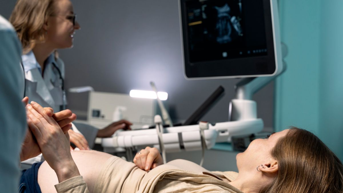
Ultrasound during pregnancy
The favorable outcome and resolution of pregnancy depends on the management tactics. Prenatal monitoring is based on an algorithm that is developed individually for each woman. The algorithm is based on the ideas of the minimum number of ultrasounds in pregnancy, the timing of which falls on each of the semesters.
Ultrasound is the main method of diagnostics for women during gestation. Future moms are interested in how often you can do ultrasound for pregnant women. The frequency of examination is appointed by the attending physician, taking into account the indications. Unlike other diagnostic techniques, ultrasound examination is safe for the expectant mother and fetus. The examination does not affect the development of the fetus and the condition of the pregnant woman. Other advantages:
Painless, non-invasive procedure.
Absence of contraindications.
High informativeness.
Availability and high speed of obtaining the result.
Ultrasound has been widely used in medicine since the 30s of the XX century. Today, ultrasound diagnostic equipment is a mandatory element of the equipment of the women's consultation and obstetric department. The question of whether you can often do ultrasound pregnant women, remains open. Scientific research on the safety of the diagnostic method for the future mother and child continues.
Why do ultrasound diagnostics
The examination helps to detect placental insufficiency, congenital pathologies and hereditary diseases in the fetus. Ask your doctor at what terms to do ultrasound to learn about possible violations in time. For more than 100 years of history of application of the method, echographic markers that indicate the presence of abnormalities in the development of the child have been studied. What can be seen during the study:
cysts in the area of the vascular plexus;
ventriculomegaly - enlargement of the brain ventricles;
excessive size of the cervical fold;
pyeloectasia - enlargement of the renal pelvis;
hyperechogenic bowel, which is often associated with intrauterine developmental delay, chromosomal disease, or intrauterine infection;
hyperechogenic focus in the ventricles of the heart, which may indicate a change in the number or structure of chromosomes.
The result of ultrasound in pregnancy shows how high the probability of congenital diseases in the child. Echographic markers are predominantly manifested in the late stages, when the gestational age is more than 14 weeks. The reliability and accuracy of the results depend on the competence of the doctor and the quality of the equipment. Modern ultrasound machines from leading manufacturers give a detailed picture of the state of different structures and organs of the fetus.
Timing of the diagnosis
The doctor who observes the patient will individually tell you when the first ultrasound of a pregnant woman is done. Usually, routine examination throughout the gestation period is carried out three times. The first examination is scheduled at the term of 12-14 weeks. What shows the diagnosis in the first trimester:
The exact gestational age.
Multiple or normal pregnancy.
Compliance of the size and structure of fetal tissues with normal indicators.
The first examination is done no earlier than 3-5 weeks, when the fetal egg is clearly visible. In the first trimester, an ectopic pregnancy is detected. Pathology is detected starting from 5-6 weeks. The fetal egg develops in the fallopian tube, not reaching the uterus. The growth of the embryo leads to the rupture of the fallopian tube and the development of a condition that is dangerous to the health and life of the woman.
Identifying a frozen pregnancy is one of the challenges of early ultrasound diagnosis. The embryo's heart beats starting in the 5th week. It is possible to determine the heartbeat during ultrasound, starting from 6-7 weeks. A frozen pregnancy is characterized by the absence of a heartbeat and fetal arrest, usually ends in miscarriage.
When ultrasounds are made in early pregnancy, the doctor pays special attention to the length of the nasal septum and the thickness of the collar zone in the fetus. To determine the gestational age, the fetal egg is measured. The method is effective at the earliest stage, when the fetus is not yet visualized. To determine the term as accurately as possible, the distance from the coccyx to the cephalic end is measured. In the second month of gestation, the diameter of the yolk sac is determined. When the embryo is 6 weeks old, the diameter is 4-5 mm. By the end of the first trimester, the development is reversed.
If the size of the yolk sac is more than 6 mm after the 10th week of gestation or less than 2 mm between the 8th and 10th week, there is a high probability of spontaneous miscarriage. Another possible complication is an unintended pregnancy.
Such results force the doctor to prescribe additional diagnostics. At what weeks do ultrasound during pregnancy, depends on the nature and dynamics of the development of disorders. Abnormal sizes of the yolk sac are among the unfavorable prognostic criteria. However, the irregular shape and size can accompany the normal course of pregnancy. To control the formation of the embryo, the doctor prescribes additional diagnostic procedures. The doctor decides how many ultrasound sessions will be needed in the end.
Pregnancy ultrasound by week: II trimester
To find out at what terms the ultrasound is done, you need to make an appointment for a consultation with a doctor. The second scheduled examination is carried out in the period from 20 to 24 weeks. What it shows:
Malformations and abnormalities in the development of the fetus.
Pathological changes in the blood supply system of the uterus.
The place of attachment of the placenta.
Pregnant women often want to know the sex of the future child, which can be determined starting from the 16th week. In practice, examinations to determine the sex of the future child are carried out starting from 22-23 weeks. Diagnosis in the early stages is associated with difficulties, for example, the position of the embryo, in which it is impossible to consider the sex characteristics.
Ultrasound of pregnancy by week: III trimester
The doctor will tell you when to do an ultrasound in pregnancy in the third trimester. The third examination according to the plan usually takes place in the period from 32 to 34 weeks. Diagnosis allows you to confirm or refute suspicions of diseases and developmental anomalies. What shows the third scheduled ultrasound in pregnancy:
The volume of amniotic fluid.
The size of the structures and organs of the fetus.
Photo of the future child.
During the examination, the doctor determines the exact date of delivery, decides on the method of delivery - natural childbirth or cesarean section.
Additional examination
In addition to routine examinations, additional screening is often carried out. If there are indications, the doctor will prescribe at what weeks to do ultrasound additionally. Unscheduled diagnosis is indicated in cases:
Aggravated heredity - the presence of close relatives who suffered from diseases that are inherited at the level of genes.
Chronic diseases diagnosed in the mother, which can affect the formation and development of the fetus.
The age of the woman exceeds 35 years. The older the woman in labor, the higher the risk of complications during gestation.
Aggravated course of pregnancy in the anamnesis. If a pregnant woman has previously experienced problems with carrying the fetus (miscarriages, frozen pregnancy), additional monitoring is required.
Adverse effects on the expectant mother. Among the negative factors - intoxication, poisoning with salts of heavy metals and other harmful substances, radioactive exposure, forced intake of pharmaceuticals during gestation.
Deviations detected during the previous screening. If in the course of the previous examination violations are detected, a repeat screening is appointed to observe the state in dynamics.
How many times to do ultrasound in pregnancy depends on the state of the future mother and fetus. In the case of therapeutic procedures, additional examination helps to monitor the effectiveness and results of therapy.
During gestation, the fertilized cell undergoes a cycle of transformations and takes the form of a person. Ultrasound of pregnancy by week is done to monitor the condition of the fetus and timely carry out therapeutic measures. Based on the results of diagnosis, the doctor predicts possible complications.
What do ultrasound in pregnancy
The doctor individually determines what ultrasound is done in pregnancy in each case. The main types of ultrasound - transvaginal and transabdominal. In the first case, the sensor is placed inside the vagina, in the second - the sensor is moved along the surface of the abdomen. Transvaginal insertion of the transducer provides better visualization. This method is usually used for the first examination when the gestational age is less than 1 month. Repeated examinations after 8-10 weeks of gestation are carried out transabdominally, so as not to provoke undesirable consequences - premature labor or miscarriage.
Where is better to do ultrasound in pregnancy
To know where it is better to do ultrasound in pregnancy, you should take into account the factors that affect the quality of diagnosis. The accuracy of the study depends on the professionalism of the diagnostician and the type of equipment. If the clinic uses advanced ultrasound machines with advanced functionality, the doctor is guaranteed to get accurate and complete results. Modern ultrasound machines form on the screen a three-dimensional 3D and four-dimensional 4D image, which significantly increases the informativeness. In 3D format, you can see the smallest details of the baby's face.
Safety and features of the procedure
Modern data confirm the safety of the procedure for the future child and mother. The technique has been used for several decades. During this period, there are no signs of negative effects of the examination on the fetus and the woman's body. There are no restrictions on the number of procedures. An additional advantage - no need for preparation. All that is needed for ultrasound in pregnancy - hygienic treatment of the study area.


