Ultrasound of pelvic organs - when it is done and indications
Ultrasound examination occupies a leading position in the diagnosis of diseases of the genital sphere in men and women. The advantages of the technique include the absence of contraindications and radiation exposure, increased informativeness, safety and accessibility. Ultrasound of the pelvic organ - painless, non-invasive procedure, which does not cause discomfort to the patient. Regular ultrasound examinations help to timely detect and effectively treat diseases.
Examination in the format of ultrasound - a safe way to obtain information, suitable for pregnant women. To do ultrasound pelvic ultrasound in men and women use ultrasound diagnostic devices. To find out how much the procedure costs, you should make an appointment for a consultation with a doctor - gynecologist, urologist.
Ultrasound of the internal genital organs: indications
Ultrasound of the internal genital organs is prescribed in case of painful sensations in the lower abdomen, discharge, bleeding and other signs of pathology. Scheduled preventive diagnostics is carried out at least once a year. Ultrasound screening in women affects the organs and structures of the reproductive system. What is included in the reproductive system:
- ovaries;
- uterus;
- cervix;
- fallopian tubes;
- vagina;
- endometrium.
At the same time, the doctor examines the bladder and the posterior space behind the uterine region. If indicated, the doctor examines the condition of regional lymph nodes, vessels and organs located in the abdominal cavity. Indications for the procedure:
- Pregnancy. Diagnosis helps to monitor the development of the fetus and the condition of the mother. Screening during pregnancy is carried out at least 3 times - 1 time in each of the trimesters. If there are indications, the doctor prescribes additional examinations.
- Menstrual disorders, including irregular, painful menstruation, bloody discharge in the middle of the cycle.
- Infertility. Examination helps to find out the causes of infertility, diagnose inflammatory processes and diseases that prevent conception.
- Suspicion of pathologies of organs that are part of the urogenital system - endometriosis, polyps, neoplasms.
- Aggravated heredity. Ultrasound screening is assigned to women whose close relatives suffered from diseases of the organs of the reproductive system. For example, a risk factor for the development of uterine myoma or ovarian cysts - genetic predisposition.
- Absence of menstruation in patients over 15 years old.
Ultrasound diagnosis with a periodicity of 1 time during the year is indicated for women who have an intrauterine device installed. Regular ultrasound monitoring is carried out for women who take hormonal contraceptives on a permanent basis.
How to perform pelvic ultrasound in women
Ultrasound examination of women is carried out in the first week of the cycle - on the 5-7th day from the beginning of menstruation. The doctor often appoints a repeat study in case of unclear diagnosis. Before learning how to do pelvic ultrasound for women, it is worth clarifying the indications for the procedure:
- Preventive examination in order to identify possible disorders and pathological changes.
- Monitoring ovulation to assess the activity of the ovaries and uterus according to the phases of the cycle.
- Diagnosis of pregnancy at the term of 3-5 weeks, monitoring the condition and development of the fetus.
- Location of intrauterine contraception - spirals.
With the help of ultrasound screening detect diseases and pathologies at an early stage - tumors and cystic formations of the ovaries, neoplasms in the bladder, ducts and intestines. Ultrasound of the uterus helps the doctor diagnose myoma and uterine fibroids, endometriosis, polyps, endometrial hyperplasia, inflammatory processes.
Examination in women is performed mainly on transabdominal or transvaginal access. In the first case, the doctor moves the sensor along the surface of the abdomen. In transvaginal examination, the sensor is placed inside the vagina. The second method is characterized by better informativeness. Abdominal ultrasound allows you to determine the location of organs, detect the presence of pathological changes - the structure and size of the pathological focus. Advantages of the technique:
- No restrictions, the possibility of conducting an examination in all patients, including children.
- Panoramic view of the organs located in the pelvis.
- Absolute non-invasiveness.
Disadvantages of the method - the need for preparation, including filling the bladder and diet for 3-5 days before the examination. Filled bladder presses on the surrounding structures, which leads to poor visualization. Factors affecting image quality are intestinal peristalsis and abdominal wall thickness. The adhesion process in the area of the study makes diagnosis more difficult.
Transvaginal method allows you to get a clear, detailed picture of individual structures located in the lower part of the abdominal cavity. The quality of the image is higher due to the absence of interference and obstacles. Another advantage of the technique is the absence of the need for preparation. To perform the diagnosis does not require filling the bladder and compliance with the diet.
The doctor determines how to do ultrasound, taking into account the age and condition of the patient. Girls and girls who are not sexually active, make diagnostics transabdominal access. In late pregnancy, the examination is carried out exclusively using the transabdominal method, so as not to provoke premature labor and miscarriage. In the absence of pregnancy and contraindications, the procedure is more often performed transvaginally. The combination of transvaginal and transabdominal techniques allows you to accurately assess the state of the genitourinary system of the patient. How to do ultrasound of the small pelvis transvaginal method:
- The patient lies down on the couch.
- The doctor introduces the sensor into the vagina, having previously put on it a gynecological condom.
- The operator moves the sensor inside the vagina to get a better look at certain areas.
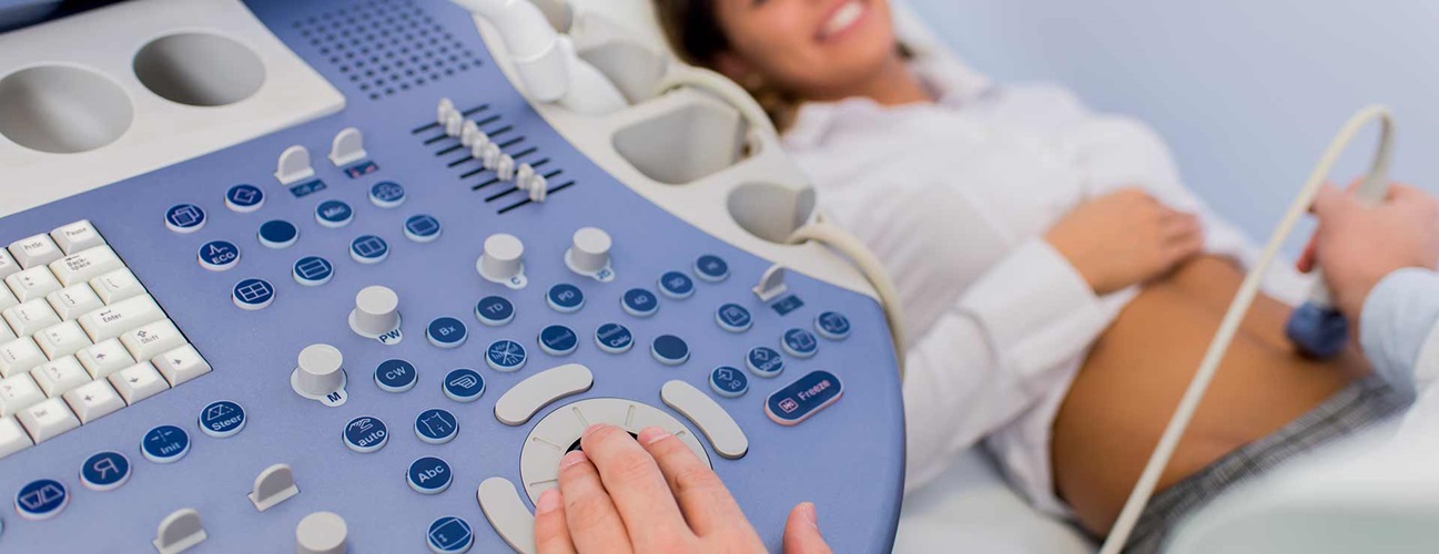
The duration of the procedure is 15-20 minutes. Transabdominal examination is carried out in the supine position. The doctor treats the surface of the abdomen with a gel that improves the conductivity of ultrasound waves. The doctor moves the transducer in different positions to see the necessary internal structures and assess their condition. Modern ultrasound machines allow you to take pictures that simplify the process of diagnosis, provide the possibility of remote consilium.
Regular diagnosis of the anatomical structure and functional state of the structures located in the lower abdominal cavity - a way to maintain health. Pelvic organs in women:
- Reproductive - uterus, ovaries, vagina, fallopian tubes.
- Digestive - parts of the intestines (sigmoid, cecum, rectum, appendix).
- Urinary - ureters, bladder, urethra.
During ultrasound screening, the doctor assesses the size, condition, morphological structure of the uterus and ovaries, reveals the number, size and localization of myoma nodes, features of endometrial transformation during the cycle. To perform ultrasound diagnostics use ultrasound machines. Modern models are characterized by an expanded set of functions and accuracy of research. Manufacturers offer the option of visualizing small details, increasing the angle of view, automatic analysis of information with the help of intelligent software. Advanced ultrasound machines provide clear images with improved contrast and high resolution, simplify the work of medical personnel and reduce the likelihood of errors due to human error.
Preparation for pelvic ultrasound
The pelvic organs of a woman throughout her life change in structure and functional activity. Childbirth and aging processes often provoke pathological changes and prolapse of organs located in the pelvis. Such violations are detected during ultrasound screening. The doctor chooses the date taking into account the indications for examination:
- 5-7 day of the cycle. Diagnosis shows the condition and pathological changes in the uterus and ovaries.
- 5-10 or 16-20 days of the cycle. The examination is carried out if the doctor suspects uterine myoma.
- 25-28 days of the cycle. Ultrasound is done if the symptoms indicate endometriosis.
- 8-10, 15-16, 23-24 days of the cycle. Monitoring the development and maturation of follicles.
The specifics of preparation depend on how the ultrasound diagnosis is carried out. For transvaginal screening, preparation is not required. If it is planned to use the transabdominal method, it is necessary to follow the rules:
- For 3-5 days before the examination, adhere to the diet - refuse to eat foods that cause flatulence (legumes, fresh vegetables, high-fat dairy products, black bread).
- Take Espumizan for two days before the ultrasound. The recommended dosage is 2 capsules three times during the day.
- Drink 1 liter of water, not containing gas, one hour before the screening.
Whether you can eat before pelvic ultrasound, will tell the doctor at a preliminary consultation. Usually the procedure is carried out on an empty stomach. If the diagnosis is scheduled for the second half of the day, it is allowed not to have a dense breakfast. Allergies to latex should be reported to the doctor in advance, if transvaginal screening is planned.
Pelvic ultrasound in men
Ultrasound of the pelvic organs for men is carried out as part of a preventive examination, if the doctor suspects diseases of the genitourinary sphere. Screening is safe for the patient and is characterized by high informativeness. Indications for ultrasonic diagnostics in men:
- Violations of the function of urination.
- Pain, discomfort, difficulties during urination.
- Insufficiently complete emptying of the bladder.
- False, imperative (not tolerating objection) urges to urinate.
- Pulling pain in the lower abdomen and perineal area.
- Painful sensations in the lumbar area, regular symptoms in the form of renal colic.
- Painful sensations in the organs of the reproductive system, including the prostate gland and scrotum.
- Discharge - purulent or bloody from the urethra.
- Inclusions of pus and blood in the ejaculate and urine.
- Unpleasant smell, atypical color of urine and ejaculate.
- Traumatic injury to the genitals, lower abdomen and bladder.
- Erectile dysfunction.
Indication for ultrasonic diagnostics is suspicion of diseases, including neoplasms in the field of reproductive and urinary organs. Diagnosis is performed in preparation for surgical intervention on organs located in the pelvis. What shows ultrasound of the small pelvis in men:
- The state, size, morphological structure of the prostate gland, surrounding soft tissues and nodes of the lymphatic system.
- The structure, size of the bladder, patency of the ureters, the volume of remaining urine.
- The structure of the seminal vesicles.
- Inflammation in the organs that make up the genitourinary system (prostatitis, cystitis).
- New growths in the area of structures located in the groin and lower abdomen.
- Concretions in the urinary tract and bladder.
- The state of the seminal ducts.
With the help of ultrasound diagnosis, the causes of erectile dysfunction, discomfort and pain, infertility are identified. This is a mandatory procedure when prostate adenoma is suspected. Ultrasound screening for preventive purposes is carried out for all men over 45 years of age with a frequency of 1 time during the year.
Ultrasound screening is done transabdominally or transrectally. Transabdominal examination is performed superficially through the wall of the peritoneum in two stages. At the first stage, the doctor views the organs with a filled bladder. Then offers the patient to urinate. At the second stage of diagnosis, the volume of urine that remains in the bladder is determined.
For research with the transrectal method, the sensor is placed in the anus and rectum. Preparation for ultrasound of the small pelvis transrectal method in men includes emptying the bowel with an enema.


