MRI of the brain - how and why it is performed, diagnostics
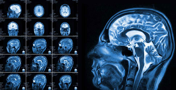
A brain MRI is a painless diagnostic procedure that produces high-quality images of a patient's head structures. Doctors use the data obtained after a brain MRI to diagnose and monitor various conditions and diseases that affect the brain or other structures of the head.
MRI of the head and brain
A head MRI is a non-invasive, painless test that uses magnetic and radio wave technology to scan the head and produce images of structures inside the skull. An MRI of the brain shows doctors conditions such as:
bleeding;
swelling;
problems with brain development;
tumors;
infections, inflammation;
damage from trauma or stroke;
problems with the blood vessels in the head.
During an MRI, the patient's head is also examined for the cause of headaches or seizures. Currently, this type of examination is the most effective and informative method of brain imaging compared to other imaging methods such as CT scans or X-rays.

What an MRI of the brain provides
MRI shows structures inside the head, including:
the brain itself;
blood vessels;
the skull and facial bones;
the structures of the inner ear;
the eyes and surrounding tissues, such as the optic nerves;
the major nerves in the head;
surrounding soft tissues and structures of the skull, such as fat, bone, muscle, and connective tissue.
MRI findings in the brain can detect abnormalities such as:
structural problems;
changes in the density of structures in certain areas, which may indicate neoplasms and cysts;
white matter disease.
MRI of the brain with contrast
Sometimes patients are prescribed contrast MRI of the brain. What does this study show? Contrast material, changes the magnetic properties of nearby water molecules, which increases the clarity of the images. The use of contrast material helps to see and evaluate more accurately:
tumors;
inflammation;
blood supply to certain areas;
the condition of the head's circulatory system.
Contrast MRI of the brain reveals what? Multiple sclerosis, stroke, dementia, and infection.
The contrast is administered through an intravenous catheter before the procedure. It is a relatively safe drug and allergic reactions are extremely rare.
When MRI of the brain is done
Doctors perform the study for a variety of reasons, including diagnosing different conditions, monitoring treatment, evaluating structural abnormalities and brain damage after a head injury.
Some of these conditions are:
A blood clot in the brain;
brain aneurysm;
stroke;
encephalitis;
epilepsy;
cysts;
dementia;
hydrocephalus;
pituitary adenoma;
brain malformations.
Indications for the examination include signs and symptoms such as:
Migraines and/or chronic headaches.
Seizures, seizures.
Frequent episodes of dizziness.
Deafness for unexplained reasons.
Vision loss without ophthalmologic cause.
Hormonal imbalances related to the hypothalamus and/or pituitary gland.
Significant changes in the patient's thinking and behavior.
Medical professionals also use MRI scans of the brain and head before head surgeries to better prepare for surgery. They also use the scans to make sure healing after surgery is going well.
How an MRI of the brain works
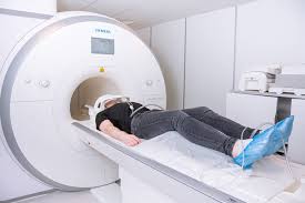
When you know what an MRI of the head is, let's tell you how the study takes place. It is carried out by a doctor who has undergone special training. Equipment - a special MRI machine, resembling a tube or a canopy with a couch. Modern MRI machines can be purchased in the company Rad Farm. We perform delivery, setup of equipment, if necessary, train staff on the operation of the technique.
To examine the brain, the patient is invited to take a seat on the couch, having previously removed all metal jewelry and accessories. The examination time is 30-60 minutes. During this time, it is necessary to remain immobile. Patients who find it difficult not to move for a long time, such as children, are examined under sedation.
The technologist performs the exam while working at a computer outside the room. He communicates with the patient through an intercom system. As a result of the scan, the doctor will receive several different types of images that are used to diagnose different conditions. During the procedure, the patient will hear loud noises that occur when the technique is operating.
Contraindications
Do not perform MRI for patients with metal-containing structures in the body, such as implants, plates, pacemakers. The procedure is contraindicated in pregnant women.


