MRI of the pituitary gland - how and why it is performed, diagnostics
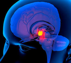
The pituitary gland is a small oval endocrine gland in our body, also known as the "master gland" because it regulates the function of other glands in our body. It secretes various hormones into the bloodstream. It consists of two lobes namely anterior lobe and posterior lobe.
The hormones secreted by the anterior lobe include:
TTG (thyroid hormone);
prolactin;
ACTH (adrenocorticotropic hormone);
growth hormone;
FSH (follicle-stimulating hormone);
LH (luteinizing hormone);
melanocyte-stimulating hormone;
enkephalins;
endorphins.
Hormones secreted by the posterior lobe of the pituitary gland include:
Oxytocin;
ADH, also known as vasopressin.
MRI of the pituitary gland is an examination of the pituitary gland to detect any abnormalities of the organ such as inflammation, overactivity or atrophy, and tumors. MRI examination of the pituitary gland provides clear images of soft tissue and helps the doctor analyze the body's tissue types and distinguish normal, functional tissues from abnormal structures.
What an MRI of the pituitary gland represents
A typical targeted pituitary MRI is imaging that produces coronal and sagittal postcontrast T1 and small-field T1 images, as well as coronal images with dynamic contrast, which are critical for identifying small microadenomas. T2-weighted sequences are also often included.

Examination of the pituitary gland assists in the evaluation and investigation:
The pituitary gland, including the funnel and posterior lobe of the pituitary gland, as well as adjacent areas;
the territory of the cavernous sinus and Meckel's cave;
the optic nerves, optic junction, and optic tracts;
the internal carotid arteries and their branches;
the diaphragm of the Turkish saddle and the borders of the pituitary fossa.
Currently, MRI of the brain or pituitary gland is considered the preferred diagnostic method for evaluating the pituitary gland, as it provides an opportunity to study in detail intracranial structures that are inaccessible to other imaging methods. Modern MRI machines for diagnostics are offered by Rad Farm.
Evaluation of pituitary gland on MRI
The pituitary gland on MRI consists of an anterior and posterior part. The anterior portion is isointense on both T1 and T2-weighted images. The posterior lobe of the pituitary gland has intrinsic high T1 signal and hypointense signal on T2-weighted images. During dynamic contrast, contrast can be seen to penetrate the gland from the funnel and gradually spread to the peripheral parts of the gland.
Age-related changes
The volume of the pituitary gland varies with hormonal status, and what would be a normal gland for one demographic would be completely abnormal for another.
Younger patients have a larger gland than people of retirement age. During puberty and pregnancy, the gland is largest. It completely fills the pituitary fossa and even protrudes beyond its borders. In the elderly, the gland is thinner, lies at the bottom of the saddle, and the pituitary fossa looks "empty".
Although one should always be cautious about measurements, they can serve to quantify what might otherwise appear to be an overly subjective impression. These are maximum values for gland height:
Children under 12 years of age: 0.6 cm, the gland is flat or slightly concave;
adolescents: 1 cm, the gland is convex, brighter in girls;
young people: in men: 0.8 cm, in women: 0.9 cm, in pregnant women: 1.2 cm;
patients over 50 years of age: there is a gradual decrease in the size of the gland.
Pituitary masses
The pituitary tumor on MRI is clearly visible, its location, morphology is identified.
MRI procedure
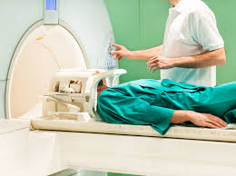
The MRI procedure of the pituitary gland takes 30-60 minutes. The MRI machine is a long tube with holes at both ends. There is also a couch that slides into the tube. If you are claustrophobic, let your doctor know. The MRI machine makes whistling or banging noises while it is taking pictures. These sounds may be loud, but they are not dangerous. They mean that the MRI machine is taking pictures.
When diagnosing pathologies of the pituitary gland, the examination is often carried out with contrast. In this case, the patient is preliminarily injected with a dye intravenously. In more detail about how MRI of the head (pituitary gland) is carried out, the doctor will tell you at a preliminary consultation.
Contraindications
Patients with metal structures in the body, such as cardiac and neurostimulators, pins, plates, artificial joints, stents can not undergo MRI because of the magnetic effects inside the device and the risk of displacement of the structure. Pregnant women are advised to refuse MRI, if the condition is not life-threatening, until delivery. For these groups of patients, the doctor will tell them how to check the pituitary gland in other ways, such as through laboratory tests for hormones.


