Computed tomography - what it is, what diseases it diagnoses, how to prepare
CT – is an informative diagnostic method that is based on the use of X-ray radiation. Non-invasive examination allows you to obtain layer-by-layer images of tissues. The software integrates slice images into 3 D graphics. Benefits of CT:
Painlessness. The examination does not cause discomfort.
High precision. During the procedure, pathologies are identified in the early stages of the preclinical course.
Universality. The technique is suitable for examining bone structures, internal organs, blood vessels, and soft tissues.
Computed tomography is considered more informative compared to MRI when examining lungs and blood vessels. CT information content within the diagnosis of tuberculosis, lung cancer and other neoplasms is 20% higher than that of MRI. Unlike a conventional radiographic examination, CT provides a detailed picture, which simplifies diagnosis. Native CT is less sensitive to study area displacement during patient movement when compared to MRI.
Compared to the radiograph, CT images show better differences between tissues of different densities. Additional plus – high speed of obtaining results. The method is suitable for diagnosing pathologies in emergency cases, which include accidents, emergencies, and suspected internal bleeding. Used to examine patients with implanted medical devices and devices.
What is computed tomography
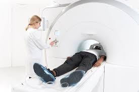
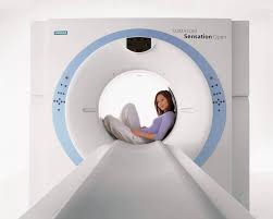
People who are interested in accurate diagnosis know what computed tomography is. Unlike X-rays, CT produces a spatial, three-dimensional image. To carry out the procedure, special equipment – tomographs is used. The X-ray source is presented in the shape of a circle, inside of which there is a mechanized table for the patient. The circle rotates around the patient's body.
At the same time, the scanning surface with 4-64 detectors takes high-resolution images. Thanks to the high image resolution, the diagnosis of heart disease and visualization of organs located in the abdominal cavity are simplified. The images enter the computer, where they are combined and transformed into a three-dimensional picture. A detailed three-dimensional image is built from two-dimensional tomograms.
Modern tomographs with intelligent functions and special software independently produce CT results with a probable diagnosis, simplifying the doctor’s work and reducing the likelihood of medical error.

Computed tomography: types
CT is native – without contrast and using contrast media. The main difference between – procedures is the introduction in the second case of special drugs into the patient’s bloodstream that change the color of physiological fluids. Computed tomography with contrast and without – the difference is the quality of imaging. The native procedure is carried out if cerebral hemorrhages, the presence of stones in the organs of the urinary system, and nodes in the lungs are suspected. Helps diagnose bone fractures and musculoskeletal diseases.
Diagnosis with contrast is necessary for better visualization of foci of infections, neoplasms, inflammatory processes and traumatic damage to soft tissues. CT with contrast – is the first choice method when assessing the condition of the vascular bed. The procedure is carried out if pulmonary embolism, dissection and aortic aneurysm are suspected. Other differences between CT with and without contrast lie in the duration of the study, the features of preparation and contraindications. Diagnosis with contrast takes longer. In the case of contrast CT, a blood test for creatinine – is a mandatory stage of preparation. Other pre-CT tests with contrast include a blood test for urea concentration and GFR.
Computed tomography: how to prepare
The specifics of preparation depend on the research site. To study the digestive tract and organs located in the abdominal cavity, you must follow a diet for 1-2 days before diagnosis. The doctor may prescribe Nosch-pa and Espumizan. To prepare for the procedure, you need to give up food 8-10 hours before the session.
Computed tomography: readings
CT scan with contrast and without the use of contrast agent is carried out to identify diseases of the musculoskeletal system, nervous, cardiovascular, and respiratory systems. The procedure is the first choice to control the performance of targeted biopsies and minimally invasive interventions. Suitable for studying the structure of the brain, spine, abdominal and chest organs. Using CT diagnostics, kidney stones and neoplasms ranging in size from 5 mm are identified. Computed tomography is performed before surgery to plan the intervention.
Computed tomography: contraindications
Patients receive a dose of radiation, which increases the likelihood of developing a malignant tumor. Due to X-rays, CT is contraindicated in pregnant women and children under 5 years of age. However, the effectiveness of diagnosis outweighs the possible risks of the procedure. Contraindications to computed tomography:
thyroid pathologies;
serious condition of the patient;
myeloma;
body weight greater than 150 kg;
recently underwent radiation therapy or radiographic diagnosis;
severe heart disease.
Contraindications to CT with contrast include childhood under 12 years of age, pregnancy and breastfeeding, allergy to contrast agent, renal failure, bronchial asthma, diabetes mellitus in the decompensation stage.


